
Electrophysiological mapping
Electrophysiological mapping gathers and interprets the heart's electrical signals to help locate and understand an arrhythmia. It may be part of an invasive study and guide a treatment decision. Mapping obtains and interprets information; ablation is a separate intervention. [1] [3]
How mapping works
During a catheter-based study, electrodes record signals in different areas of the heart. The team compares the signals and their locations to investigate where an arrhythmia starts or how it continues. Controlled stimulation and other maneuvers may help clarify the mechanism. [1] [3]
Mapping can help in selected fast heart rhythms and procedures for different arrhythmias. The need for mapping, the technique and possible treatment depend on the assessment and the clinical question. Not every study requires a three-dimensional system. [1]
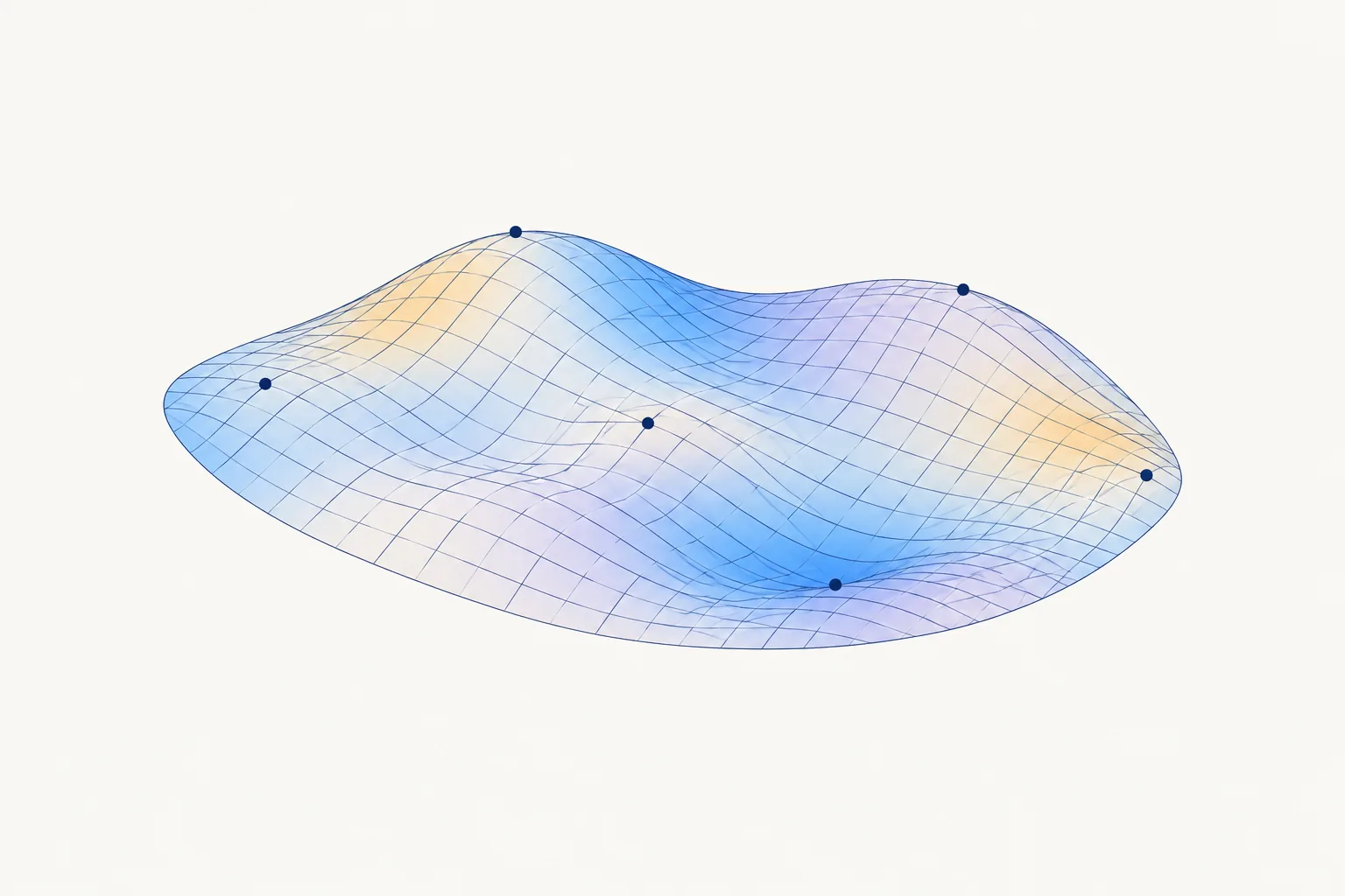
What is a 3D electroanatomical map?
In some cases, a system combines catheter positions with recorded signals to create a three-dimensional reconstruction of the area being studied. It is a representation produced by the system, not a photograph of the inside of the heart. [1]
An activation map organizes signal timing relative to a reference and helps show how electrical activity travels. A voltage map shows the amplitude of signals recorded at different points. They answer different questions and may be considered together, depending on the case. [1] [2]
Understanding colors and limitations
Colors depend on the type of map, scale and chosen settings. They are not a universal diagnostic code and do not by themselves demonstrate the result of an ablation. Interpretation considers the signals, catheter contact, rhythm and area actually examined. [1] [2]
Low-voltage signals may raise questions about the tissue in certain contexts, but they do not prove fibrosis on their own. The map may be incomplete or require further interpretation; its quality depends on the information collected. [1] [2]
Three-dimensional mapping can help reduce fluoroscopy, a type of X-ray imaging, in some procedures. This does not mean every procedure will be performed without radiation. The need for imaging depends on the case and the strategy used. [1]
Mapping and ablation are different
Mapping seeks information about electrical activity. Ablation applies energy to treat a chosen target. In some cases the map helps guide this intervention, but mapping does not mean energy will be applied or that treatment will succeed. The team discusses the indication, alternatives and consent. [1] [3]
Preparation, risks and recovery
When performed during an invasive study, mapping shares the care requirements and risks of the catheter procedure. Tell the team about medicines, including blood thinners, allergies and the possibility of pregnancy. Ask for individual instructions on fasting, medicines and an accompanying person; do not change your medicines on your own. [3] [4] [6]
Bleeding, bruising, infection or vessel injury can occur at the access site. Other risks include arrhythmia, blood clots, heart perforation with bleeding, damage to the conduction system requiring a pacemaker, heart attack and stroke; death is rare. Risks vary with the procedure and your clinical situation. If ablation or another procedure is also planned, discuss its specific additional risks. [3] [4]
After catheter removal, the heart rhythm and access site are monitored. Ask when you can drive, exercise and return to your activities; the plan depends on your recovery and any associated procedures. [3] [4]
When to seek medical help
Get immediate medical help for chest pain or tightness, shortness of breath after the procedure, sudden signs of stroke such as facial drooping, weakness on one side or trouble speaking, sudden increase in swelling at the access site, or bleeding that does not stop with firm pressure. In Brazil, call SAMU at 192; elsewhere, call your local emergency number. Do not wait for a reply from the scheduling channel. [4] [5]
Fever, drainage, increasing bruising, or a change in color or sensation in the arm or leg used for access requires immediate contact with the care team. If these signs are severe or getting worse, seek urgent care. [3] [4]
Frequently asked questions
Does all mapping use a 3D reconstruction?
No. Electrical recordings and their interpretation can be used without a three-dimensional reconstruction. The strategy depends on the arrhythmia and the procedure. [1]
Do colors show where there is fibrosis?
Not necessarily. Colors represent selected parameters and scales. A low-voltage signal can have different explanations and does not confirm fibrosis on its own. [1] [2]
Does a map mean I will have ablation?
No. A map provides information; ablation is a separate treatment. The decision depends on the case and consent. [3]
Does mapping eliminate the use of X-rays?
Not necessarily. Three-dimensional systems can reduce fluoroscopy in some procedures, but imaging and radiation use depend on the strategy and clinical situation. [1]
Understand your evaluation
Bring your test results and medication list to discuss the proposed procedure, its benefits, limitations and alternatives. The scheduling channel is not an emergency service.
Contact our teamScientific references
- APHRS/HRS/EHRA/LAHRS. Expert consensus on three-dimensional mapping systems for tachycardia, 2019.
- EHRA. Clinical consensus on atrial tachycardia, 2025.
- American Heart Association. Electrophysiology Studies.
- Mayo Clinic. EP study.
- Brazilian Ministry of Health. SAMU 192.
- Johns Hopkins Medicine. Electrophysiological Studies.