
What a pacemaker does
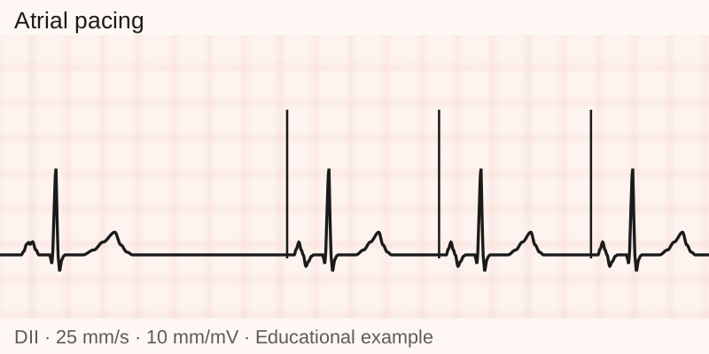
The heart has an electrical system that starts and conducts heartbeats. When this system works slowly or has blocks, fatigue, dizziness, shortness of breath and fainting may occur. The pacemaker provides the necessary pacing according to its programming.
In a conventional system, a small generator sits under the skin, usually below the collarbone, connected to leads that reach the heart through veins. Some people need occasional pacing; others need pacing most of the time. A conventional pacemaker does not deliver defibrillation shocks. [1]
Pace
- Sense
The system monitors electrical activity.
- Pace
It sends impulses according to need and programming.
When implantation is indicated
Indications include abnormalities of the sinus node, the heart's natural pacemaker, when they cause symptoms related to a slow rhythm. The device may also be indicated for certain blocks in the passage of impulses from the atria to the ventricles. Some advanced blocks may require a pacemaker even without symptoms, after reversible causes have been investigated.
The assessment seeks to relate symptoms to abnormalities recorded on the electrocardiogram and, when necessary, on a Holter monitor or longer monitoring. An echocardiogram helps assess heart structure and function.
A low heart rate alone, including during sleep or in athletes, does not establish an indication. In atrial fibrillation, the device may be needed when a slow ventricular rhythm causes symptoms or is associated with significant blocks. [1]
Pacemaker, ICD and resynchronization device: what is the difference?
A conventional pacemaker can pace one or two chambers. There are also models without leads, called leadless, implanted directly in the heart through a catheter inserted into a vein, for selected cases. The choice considers the heart's rhythm, anatomy and function.
- Pacemaker: treats certain slow rhythm and electrical conduction abnormalities.
- Defibrillator (ICD): treats potentially fatal ventricular arrhythmias. Models with leads through veins also offer pacing for slow heartbeats; a standalone subcutaneous ICD does not offer this function on a chronic basis.
- Resynchronization device (CRT): aims to improve coordination of the ventricles in selected patients. CRT-P provides pacing; CRT-D adds defibrillation. [1] [4]
How to prepare and what happens during implantation
Bring your test results and medication list. Report allergies, fever or recent infection. The team determines fasting, necessary tests and what to do with anticoagulants and antiplatelet drugs. Do not stop these medications on your own, and arrange for someone to accompany you at discharge.
Conventional implantation is performed under local anesthesia and, when indicated, sedation. Through an incision below the collarbone, the physician positions the leads through a vein using imaging guidance and connects the generator. The device is tested and programmed before discharge.
The duration of the procedure and hospital stay varies with the technique, clinical condition and recovery. [6] [3]
What are the risks?
Implantation may cause bleeding or hematoma, infection, lead displacement, thrombosis and pneumothorax, which is air entering around the lung. Cardiac perforation with blood accumulating around the heart is a potentially serious complication.
When infection involves the generator, its pocket under the skin or the leads, treatment generally requires removal of the entire system and antibiotics. A superficial incision infection without device involvement is assessed and treated differently. Fever, discharge, wound opening or progressive redness require medical assessment; do not try to decide on your own whether the infection is superficial. Lead or generator problems may also require another intervention. [10]
Recovery and follow-up
Before discharge, ask for instructions on dressings, bathing, pain control and follow-up. Limits on lifting weight, exercise and moving the arm depend on the procedure. Avoiding strenuous effort does not mean keeping the arm still: prolonged immobilization can cause shoulder pain. Follow the team's individual advice. [3]
Keep the device identification card. At follow-up visits, the team uses a device called a programmer to check the battery, leads and rhythm records and adjust pacing. Follow-up continues even when you feel well; visit frequency depends on the case. [8]
In some people, frequent right ventricular pacing may reduce the strength of heart contraction over time. The team therefore follows symptoms and heart function as well as the battery and leads. The pacing technique takes this risk into account. [11]
When available for the device and organized by the service, remote monitoring allows device data to be sent between visits. The team explains how it works and when the data are assessed. Sending data does not guarantee an immediate response or replace emergency care. [12]
Are there alternatives to a pacemaker?
Some bradycardias improve by treating their cause, such as medication effects, thyroid abnormalities or electrolyte disturbances. When safe, investigating and correcting these conditions is part of the assessment. [1]
In selected cases of severe recurrent reflex fainting with a marked fall in heart rate despite conventional measures, cardioneuroablation may be discussed by a specialist team. The decision requires confirmation of the mechanism of the fainting and discussion of benefits, risks and uncertainty about long-term outcomes.
For other bradycardias related to the vagus nerve, the international consensus considers its use investigational, in controlled studies. The technique does not replace a pacemaker indicated for disease of the heart's own electrical system. [5]
See also the guide to cardiac arrhythmia treatment.
When to seek care
Fainting, chest pain, sudden or severe shortness of breath and significant bleeding require urgent care. Go to an emergency department or call SAMU, 192.
Fever, discharge, wound opening or increasing redness require assessment the same day. Contact the team; if you cannot obtain care, go to an emergency department. New dizziness and persistent hiccups should be reported promptly to the team. Do not wait for the scheduled follow-up visit. [7] [8]
Frequently asked questions
Can someone with a pacemaker have an MRI scan?
In many cases, yes. The team must check whether the generator and leads together have a classification that allows the scan under specific manufacturer conditions, called an MRI-conditional system. Even systems without this classification may be assessed at centers with a specific protocol. The scan requires appropriate planning, programming and monitoring. Say that you have a pacemaker before scheduling the scan. [1] [2]
How long does the battery last?
The battery lasts several years, depending on the model, programming and how much pacing the heart needs. With conventional pacemakers, follow-up visits allow generator replacement to be planned while retaining the leads when they are in good condition. In leadless models, the battery and device are inside the heart: the strategy at the end of service life is different and should be planned with the team. [8] [4]
Can I use a cell phone and household appliances?
Generally, yes, following the manufacturer's instructions. Keep cell phones and objects with magnets at least 15 cm from the generator and avoid the pocket over the device. Tell healthcare professionals you have a pacemaker before tests, surgery and treatments that use electrical energy. [9]
Can I stop medication after implantation?
A pacemaker does not replace treatment for other heart diseases. Anticoagulants prescribed for atrial fibrillation, for example, may still be needed. Any adjustment before or after implantation should be determined by the physician.
When can I return to driving and work?
The time frame depends on the reason for implantation, any fainting episodes, recovery and occupation. Discuss your return at the medical assessment and follow applicable licensing rules. Arrange accompanied transport at discharge.
Discuss whether a pacemaker is indicated
For your evaluation in São Paulo, bring available test results and your medication list. Explain when symptoms occur.
Request an evaluation via WhatsApp+55 11 92137-6068 · Scheduling channel, not for emergencies.
Meet the medical teamScientific references
-
Teixeira RA et al. Diretriz Brasileira de Dispositivos Cardíacos Eletrônicos Implantáveis, 2023
Arq Bras Cardiol. 120(1):e20220892. Indications, device types and magnetic resonance imaging.
-
Glikson M et al. ESC Guidelines on cardiac pacing and cardiac resynchronization therapy, 2021
Europace. 2022;24:71-164. The 2021 guideline, including evaluation and MRI in conditional and nonconditional systems.
-
Burri H et al. EHRA consensus on pacemaker and defibrillator implantation technique, 2021
Europace. 23:983-1008. Procedure, complications and care after implantation.
-
American Heart Association. Statement on care of patients with implantable devices, 2024
Periprocedural Management and Multidisciplinary Care Pathways for Patients With Cardiac Implantable Electronic Devices. Distinguishes the functions of ICD types.
-
EHRA/HRS/APHRS/LAHRS. Statement on cardioneuroablation, 2024
Europace. 26:euae206. Selection of patients with reflex syncope and functional bradyarrhythmias.
-
NHLBI. Before getting a pacemaker
Patient guidance on preparation, medications and discharge planning.
-
NHLBI. After getting a pacemaker
Recovery, complications and signs that should be reported to the team.
-
American Heart Association. Living With Your Pacemaker
Device card, follow-up, battery and warning symptoms.
-
American Heart Association. Devices That May Interfere With ICDs and Pacemakers
Precautions for cell phones, magnets and medical procedures.
-
Blomström-Lundqvist C et al. International EHRA consensus on infection of implantable cardiac devices, 2020
Europace. 2020;22:515-549. Distinguishes superficial incision infection from system infection and recommends complete removal for confirmed infection.
-
HRS/APHRS/LAHRS. Guideline on cardiac physiologic pacing, 2023
Monitoring ventricular function in people with substantial right ventricular pacing and selecting a pacing strategy.
-
HRS/EHRA/APHRS/LAHRS. Consensus on remote monitoring of implantable cardiac devices, 2023
Organization of remote monitoring; data transmission does not replace emergency care.